Tescan LYRA-3 Dual Beam FIB/SEM for Radiological & Non-Radiological Samples
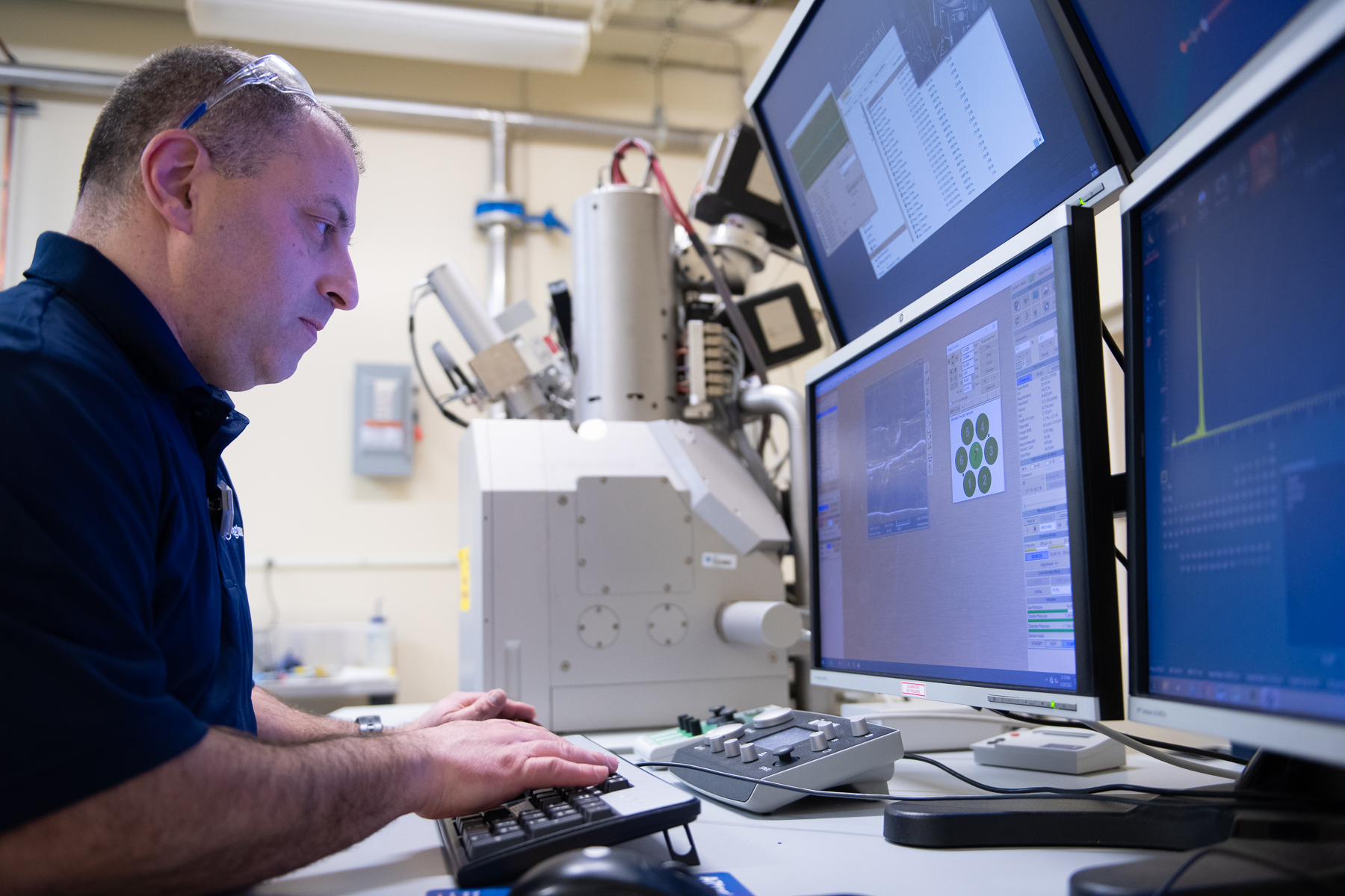
Dual beam FIB, capable of SEM imaging at variable pressures, and equipped with an Orsay Physics Canion FIB column, with XEDS and EBSD detectors.
Tescan CLARA Ultra High Resolution SEM
Advanced ultra high-resolution imaging capability (0.9 nanometer resolution), variable pressure imaging, energy filtering imaging and XEDS detector.
Transmission Electron Microscopy
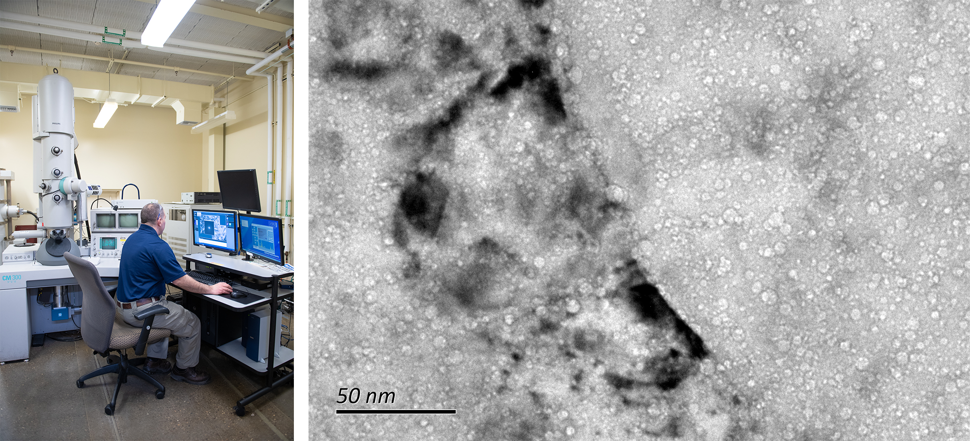
Left Image: FEI CM-300 FEG TEM/STEM equipped with Oxford X-max T80 XEDS detector, HAADF detector and Gatan Orius CCD camera. Right Image: TEM micrograph of highly neutron irradiated Alloy X-750.
Hot Cell Shielded SEM
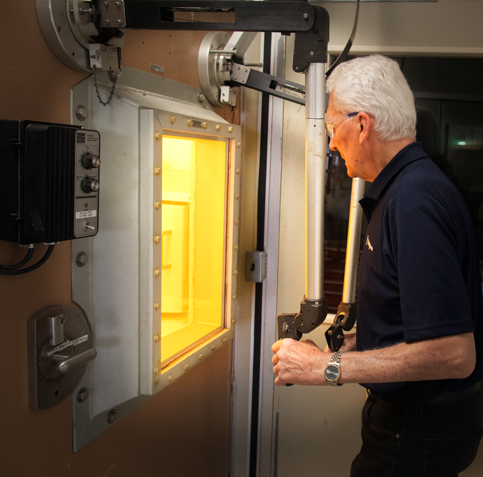
Dedicated shielded SEM (Tescan Vega-3 XMU) attached to Hot Cell is used for failure analysis of extremely radioactive components.
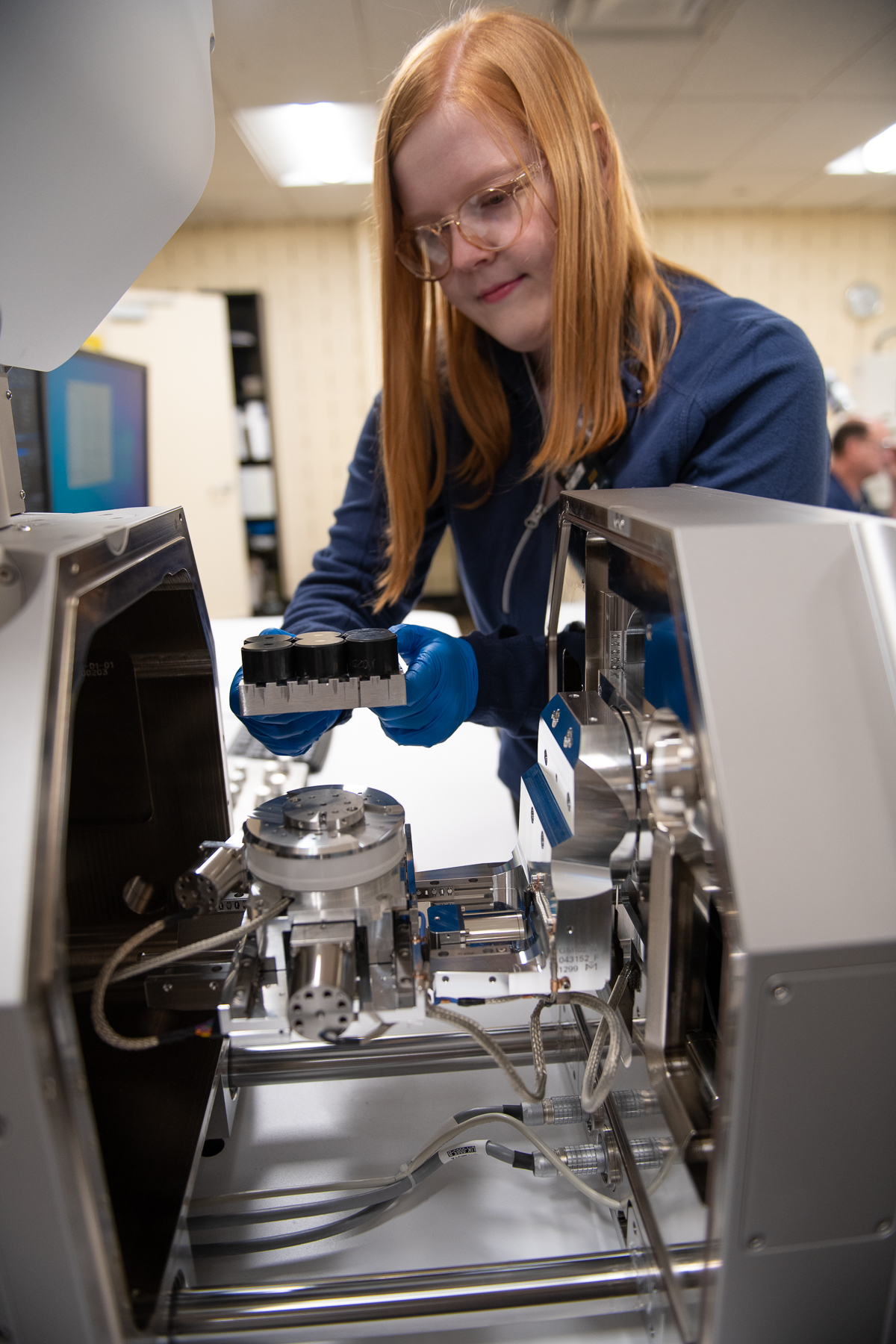
FIB sample preparation for subsequent transmission electron microscopy evaluation
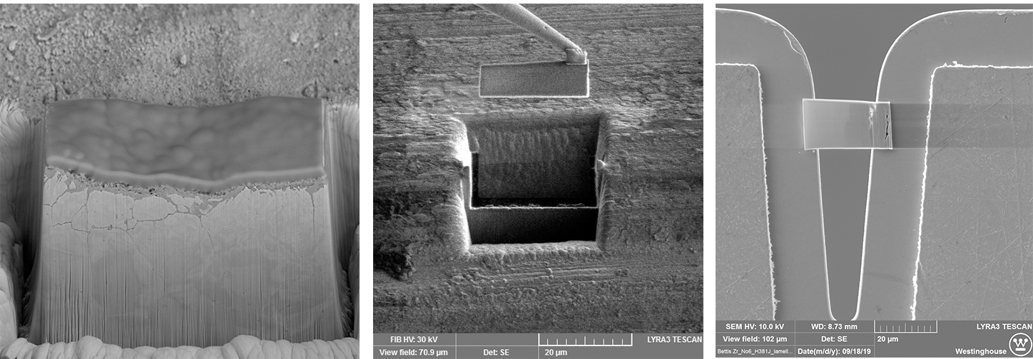
Left: An ion-milled pillar used to generate 3D tomography of oxide formed on alloy 600 steam generator tubing. Middle: In-situ extraction of a TEM lamella. Right: A TEM lamella placed on a TEM grid prior to thinning.
SEM micrographs
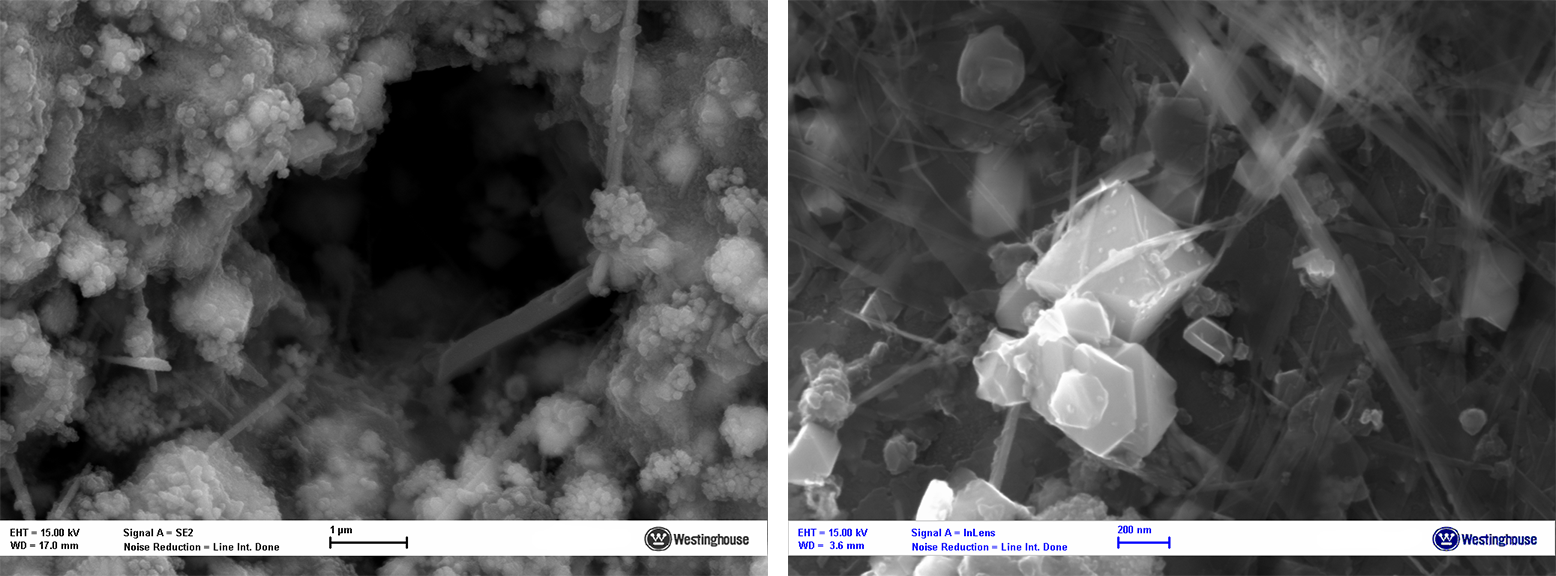
Left: High resolution image of a boiling chimney in a fuel crude flake.
Right: Oxide deposit on the ID surface of an alloy 600 steam generator tube.
Right: Oxide deposit on the ID surface of an alloy 600 steam generator tube.
1920 and up
1440 and up
1280 and up
Desktop
Tablet
Mobile Landscape
Mobile Portrait
